Body cavity
In zoology, body cavity generally refers to the space, or cavity, located between an animal’s outer covering (epidermis) and the outer lining of the gut cavity—a fluid-filled space where internal organs develop. However, the term sometimes is used synonymously with the coelom or "secondary body cavity," which is more specifically that fluid-filled body cavity between the digestive tract and the outer body wall that is completely enclosed by cells derived from mesoderm tissue in the embryo. The broadest definition of the term body cavity is any fluid-filled space in a multicellular organism, including the digestive tract.
The concept of body cavity has been important in comparative studies of the body plans used by different taxonomic groups, ranging from simple organisms with two germ layers (ectoderm and endoderm) that lack a body cavity, to organisms with three germ layers (a mesoderm also) that lack a body cavity, to those with a cavity forming between the mesoderm and endoderm and not completely lined with mesoderm, to those with a true coelom completely lined with mesoderm.
Overview
Primary and secondary body cavities, acoelomates, pseudocoelomates, and coelomates
Some animals lack any cavity; their cells are in close contact with each other, separated only by the extracellular matrix. Such organisms are known as acoelomates and have what can be called a "compact organization." However, many organisms have some type of cavity: Small interstitial spaces between cells, tube-like systems, large spaces, repeating units, and so forth (Schmidt-Rhaesa 2007).
Generally, two structural types of body cavities are recognized. One type of body cavity may be termed a primary body cavity and the other termed a secondary body cavity. More common terminology is to call one type of body cavity a pseudocoelom, and animals with this body plan pseudocoelomates, and the other type of body cavity a coelom, and animals with this body plan coelomates.
Since a cavity itself lacks features, body cavities can only be characterized on the basis of the surrounding structures or those structures internal to the cavity (Jenner 2004). A coelom is a fluid-filled body cavity that separates the digestive tract and the outer body wall and is completely lined with mesoderm (Simmons 2004). The surfaces of the coelom are covered with a peritoneum, which is a slick epithelial layer (Yeh 2002). Schmidt-Rhaesa (2007), among others, equates coelom with secondary body cavity; "the secondary body cavity is commonly named the coelom." The pseudocoelom is a fluid-filled body cavity that separates the digestive tract and the outer body wall and is not completely lined with mesoderm (Simmons 2004). This pseudocolom, which develops between the mesoderm and the endoderm, is a persistent blastocoel, or fluid-filled cavity, of the blastula stage of the embryo (Yeh 2002). Schmidt-Rhaesa (2007), among others, equates the term primary body cavity with the pseudocoelom: "The primary body cavity is sometimes called a pseudocoel." Schmidt-Rhaesa (2007), in the book The Evolution of Organs, actually differentiates the two types of cavities as the primary body cavity has an extracellular matrix that borders the entire cavity, whereas in the secondary body cavity, there is a cellular layer (epithelium) that itself rests on the extracellular matrix.
However, although coelom is unambiguously defined (Schmidt-Rhaesa 2007), the terminology of primary and secondary cavities, and aceoelomate and pseudocoelomate, although long appearing in the literature, are not rigorously defined and in some cases there has been a misleading use of the terms (Jenner 2004). For example, Jenner (2004) references the use of acoelomate also for some animals with a primary body cavity. And Yeh (2002) refers to the primary body cavity as including the digestive system (gut tube or visceral tube) and the secondary body cavity as including both organisms with a pseudocoelom or with a true coelom (for example, "animal species with a secondary body cavity, either a pseudocoelom or a true coelom"). That is, according to Yeh, acoelomates, such as sponges and flatworms, have a single body cavity, and pseudocoelomates, such as roundworms and rotifers, have a secondary body cavity. Simmons (2004) similarly notes that "primitive animals … developed only one major body cavity, the digestive tract" and "all triploblastic animals pass the Playthelminthes have some form of secondary body cavity."
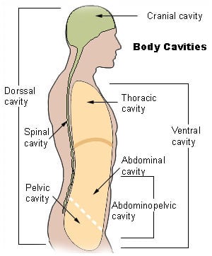
Note that the term human body cavities normally refers to the ventral body cavity, because it is by far the largest one in area.
Germ layers and coelom formation
Other than sponges, animals develop two or three germ layers during gastrulation (development of the embryo from the blatula to a gastrula). A germ layer is a layer of cells that gives rise to a specific structure in the organism, with the cells on the outside, known as the ectoderm, becoming the covering and those on the inside, known as the endoderm, becoming the gut lining (Towle 1989). Most animals form a third layer called a mesoderm, an embryonic layer that forms between the endoderm and ectoderm, and which gives rise to the muscles, skeleton, blood, blood vessels, and other interior body linings (Towle 1989).
All organisms more complex than a platyhelminthes have a coelom, whose lining is formed by the mesoderm. In deuterostomes, the mesoderm forms when there is division of the cells at the top of the gastrula; in protostomes, the cells split at the junction of the endoderm and ectoderm during gastrulation and there is rapid division of cells (Towle 1989). In coelomates, the mesodermal cells spread out and make the coelom, but in pseudocoelomates, such as the roundworm, the mesoderm lines the body cavity but does not expand to form a lining of the organs, forming rather a pseudocoelom ("false-body cavity") (Towle 1989).
Body plans
The type of body cavity places an organism into one of three basic groups according to body plan:
- Coelomate body plan. Coelomates (also known as eucoelomates—"true coelom") have a fluid-filled body cavity called a coelom with a complete lining called peritoneum derived from mesoderm (one of the three primary tissue layers). The complete mesoderm lining allows organs to be attached to each other so that they can be suspended in a particular order while still being able to move freely within the cavity. Most bilateral animals, including mollusks, annelids, arthropods, echinoderms, and all the vertebrates, are coelomates.
- Pseduocoelomate body plan. Pseudocoelomate animals have a "pseudocoel" or "pseudocoelom" (literally “false cavity”), which is a fully functional body cavity. Tissue derived from mesoderm only partly lines the fluid filled body cavity of these animals. Thus, although organs are held in place loosely, they are not as well organized as in a coelomate. All pseudocoelomates are protostomes; however, not all protostomes are pseudocoelomates. Examples of pseudocoelomates are roundworms and rotifers. Pseudocoelomate animals are also referred to as Hemocoel and Blastocoelomate.
- Acoelomate body plan. Acoelomate animals have no body cavity at all. Organs have direct contact with the epithelium. Semi-solid mesodermal tissues between the gut and body wall hold their organs in place. There are two types of acoelomate body plans. The first is characterized by two germ layers—an ectoderm and endoderm—that are not separated by a cavity, as seen in the sponges and cnidarians. The second is characterized by three germ layers—ectoderm, mesoderm, and endoderm—that are not separated by a cavity. An example of this body plan is a flatworm (Towle 1989).
Note, however, even within a particular taxonomic group, there may be cases of organisms reflecting two different body plans. Such would be the case, for example, where the larva of an organism may be a pseduocoelomate, being small and with respiration able to take place by diffusion, while the large adult organism may be a coelomate.
Coelomate body plan
A coelom is a cavity lined by an epithelium derived from mesoderm. Organs formed inside a coelom can freely move, grow, and develop independently of the body wall while fluid cushions and protects them from shocks. Arthropods and mollusks have a reduced (but still true) coelom. Their principal body cavity is the hemocoel of an open circulatory system.
Mammalian embryos develop two coelomic cavities: The intraembryonic coelom and the extraembryonic coelom (or chorionic cavity). The intraembryonic coelom is lined by somatic and splanchnic lateral plate mesoderm, while the extraembryonic coelom is lined by extraembryonic mesoderm. The intraembryonic coelom is the only cavity that persists in the mammal at term, which is why its name is often contracted to simply coelomic cavity. Subdividing the coelomic cavity into compartments, for example, the pericardial cavity, where the heart develops, simplifies discussion of the anatomies of complex animals.
Coelom formation begins in the gastrula stage. The developing digestive tube of an embryo forms as a blind pouch called the archenetron. In Protostomes, a process known as schizocoelus happens: as the archenteron initially forms, the mesoderm splits to form the coelomic cavities. In Deuterostomes, a process known as enterocoelus happens: The mesoderm buds from the walls of the archenteron and hollows to become the coelomic cavities.
Among advantages of a coelom is it allows for more extensive growth of organs, including the digestive tract, it permits the formation of an efficient circulatory system, the fluid can transport materials faster than by diffusion, there is space provided for gonads to develop during the breeding season or for young to grow in those animals, and so forth (Simmons 2004).
The evolutionary origin of the coelom is uncertain. The oldest known animal to have had a body cavity is Vernanimalcula. Current evolutionary theories include the acoelomate theory, where the coelom evolved from an acoelomate ancestor, and the enterocoel theory, where the coelom evolved from gastric pouches of cnidarian ancestors.
Pseudocoelomate body plan
In some protostomes, the embryonic blastocoele persists as a body cavity. These protostomes have a fluid-filled main body cavity unlined or partially lined with tissue derived from mesoderm. This fluid-filled space surrounding the internal organs serves several functions like distribution of nutrients and removal of waste or supporting the body as a hydrostatic skeleton.
The term pseudocoelomate is no longer considered a valid taxonomic group, since it is not monophyletic. However, it is still used as a descriptive term. A pseudocoelomate is any invertebrate animal with a three-layered body and a pseudocoel. The coelom appears to have been lost or reduced as a result of mutations in certain types of genes that affected early development. Thus, pseudocoelomates evolved from coelomates (Evers and Starr 2006).
Animals with this body plan:
- Lack a vascular blood system (diffusion and osmosis circulate nutrients and waste products throughout the body)
- Lack a skeleton (hydrostatic pressure gives the body a supportive framework that acts as a skeleton)
- Lack segmentation
- The body wall of epidermis and muscle is often syncytial and usually covered by a secreted cuticle
- Are mostly microscopic
- Include parasites of almost every form of life (although some are free living)
Examples of pseudocoelomates include:
- Nematoda (roundworms)
- Rotifera (rotifers)
- Kinorhyncha
- Nematomorpha, nematomorphs, or horsehair worms
- Gastrotricha
- Loricifera
- Priapulida
- Acanthocephala (spiny-headed worms)
- Aschelminth animals
- Entoprocta
Acoelomate body plan
Lacking a fluid-filled body cavity presents some serious disadvantages. Fluids do not compress, while the tissue surrounding the organs of these animals do. Therefore, acoelomate organs are not protected from crushing forces applied to the animal’s outer surface. There are restrictions on size and locomotion, for any increase in size would require increase in volume of tissue to be nourished, but the solid body places prevents formation of an efficient circulating system and the solid body places pressure on organs during movement (Simmons 2004).
Organisms showing acoelomate formation include the platyhelminthes (flatworms, tapeworms, and so on) These creatures do not have a need for a coelom for diffusion of gases and metabolites, as the surface area to volume ratio is large enough to allow absorption of nutrients and gas exchange by diffusion alone, due to dorso-ventral flattening.
ReferencesISBN links support NWE through referral fees
- Evers, C.A., and L. Starr. 2006. Biology:Concepts and Applications, 6th edition. Thomson. ISBN 0534462243.
- Jenner, R. A. 2004. Part II: Character evaluation. Body cavities. Contributions to Zoology 73 (1/2). Retrieved August 1, 2008.
- Schmidt-Rhaesa, A. 2007. The Evolution of Organ Systems. Oxford University Press. ISBN 0198566697.
- Simmons, K. 2004. The acoelomate-coelomate split. University of Winnipeg: Biology 05-1116-3. Retrieved August 1, 2008.
- Solomon, E.P., L.R. Berg, and D.W. Martin. 2002. Biology. Pacific Grove, Calif: Brooks/Cole. ISBN 0534391753.
- Towle, A. 1989. Modern Biology. Austin, TX: Holt, Rinehart, and Winston. ISBN 0030139198.
- Yeh, J. 2002. Body cavities. NovelGuide.com. Retrieved August 1, 2008.
Credits
New World Encyclopedia writers and editors rewrote and completed the Wikipedia article in accordance with New World Encyclopedia standards. This article abides by terms of the Creative Commons CC-by-sa 3.0 License (CC-by-sa), which may be used and disseminated with proper attribution. Credit is due under the terms of this license that can reference both the New World Encyclopedia contributors and the selfless volunteer contributors of the Wikimedia Foundation. To cite this article click here for a list of acceptable citing formats.The history of earlier contributions by wikipedians is accessible to researchers here:
The history of this article since it was imported to New World Encyclopedia:
Note: Some restrictions may apply to use of individual images which are separately licensed.

