Nervous system
The nervous system is the network of specialized cells, tissues, and organs in a multicellular animal that coordinates the body's interaction with the environment, including sensing internal and external stimuli, monitoring the organs, coordinating the activity of muscles, initiating actions, and regulating behavior. At the cellular level, the nervous system is defined by the presence of a special type of excitable cell called a neuron (or "nerve cell") that transmits impulses. All parts of the nervous system are made of nervous tissue, which contains the two main categories of cells: neurons and supporting glia cells. An example of an organ that is part of the nervous system is the brain, which serves as the center of the nervous system in all vertebrate and most invertebrate animals.
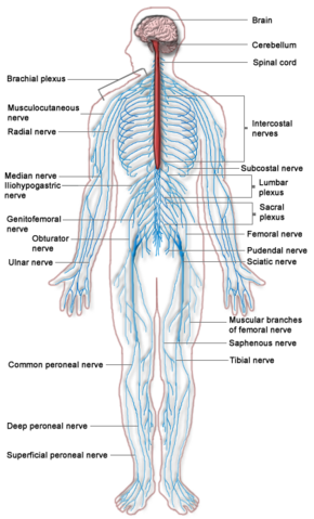
This major coordinating system is found in most invertebrates and all vertebrates, but is most complex in vertebrate animals. The only multicellular animals that have no nervous system at all are sponges, placozoans, and mesozoans, which have very simple body plans. In vertebrates, the nervous system is divided into the central nervous system (CNS), comprising the brain and spinal cord, and the peripheral nervous system (PNS), consisting of all the nerves and neurons that reside or extend outside the central nervous system, such as to serve the limbs and organs. The large majority of what are commonly called nerves (which are actually axonal processes of nerve cells) are considered to be part of the peripheral nervous system.
Cephalization is a trend seen in the history of life whereby nervous tissue in more advanced organisms is concentrated toward the anterior of the body. This process culminates in a head region with sensory organs. The human brain is the most complex known living structure, with some 86 billion nerve cells and trillions of neuronal connections; millions of information transfer processes take place in remarkable coordination every second in the human central and peripheral nervous system. There also are more than 1,000 disorders of the human brain and nervous system, with neurological disorders affecting up to one billion people worldwide. Neurology is the medical specialty dealing with disorders and diseases of the nervous system. Neuroscience is the field of science that focuses on the study of the nervous system.
At the most basic level, the function of the nervous system is to send signals from one cell to others, or from one part of the body to others. At a more integrative level, the primary function of the nervous system is to control the body, by extracting information from the environment and transmitting, processing, and acting on this information. In order for an individual to grow and develop, it needs to be continuously engaged in reciprocal relationships with its environment. Furthermore, the ubiquity of the nervous system among multicellular organisms reflects the unity in nature.
Overview
The nervous system is the part of an animal's body that coordinates the voluntary and involuntary actions of the animal and transmits signals between different parts of its body. This coordinating system derives its name from nerves, which are cylindrical bundles of fibers that emanate from the brain and central cord, and branch repeatedly to innervate every part of the body (Kandel et al. 2000). Nerves actually consist of a cable-like bundle of axons (the long, slender projection of a neuron), along with a variety of membranes that wrap around them and they are capable of transmitting electrical signals called nerve impulses or, more technically, action potentials. Nerves are large enough to have been recognized by the ancient Egyptians, Greeks, and Romans, but their internal structure was not understood until it became possible to examine them using a microscope (Finger 2000).The neurons that give rise to nerves do not lie entirely within the nerves themselves—their cell bodies reside within the brain, central cord, or peripheral ganglia (Kandel et al. 2000).
Cellular components and their functions
The nervous system contains two main categories or types of cells: neurons and glial cells.
Neurons
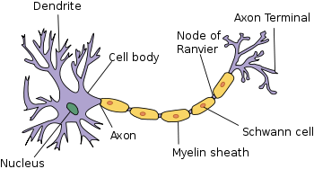
Neurons, also known as neurones and nerve cells, are electrically excitable cells that process and transmit information. Neurons have a wide variety of structures, sizes, and electrochemical properties. However, most neurons are composed of four main components:
- A soma, or cell body, is the central part of the neuron and contains the nucleus.
- Dendrites are cellular extensions with many branches, and a neuron commonly contains one or more dendritic trees that typically receive input. A dendrite may receive chemical signals from the axon termini of other neurons and convert these into small electric impulses to transmit to the soma.
- An axon is the finer, cable-like projection of the cell body that can extend tens, hundreds, or even tens of thousands of times the diameter of the soma in length. The axon is specialized for the conduction of a particular electric impulse, called the action potential, which travels away from the cell body and down the axon.
- The axon terminal refers to the small branches of the axon that form the synapses, or connections with other cells.

Neurons can be distinguished from other types of cells in a number of ways, but their basic function and most fundamental property is that they communicate with other cells via chemical or electric impulses across a synapse—the junction between cells that contains molecular machinery allowing rapid transmission of the electrical or chemical signals. Essentially, a typical process is that an electrochemical wave called an action potential (an electrical signal that is generated by utilizing the electrically excitable membrane of the neuron) is generated and this action potential travels along the axon to the synapse. There the action potential can cause the release of a small amount of neurotransmitter molecules, which bind to chemical receptor molecules located in the membrane of the target cell. A cell that receives a synaptic signal from a neuron may be excited, inhibited, or otherwise modulated. Most neurons send signals via their axons, although some types are capable of dendrite-to-dendrite communication.
Synapses may be electrical or chemical. Electrical synapses make direct electrical connections between neurons (Hormuzdi et al. 2004), but chemical synapses are much more common, and much more diverse in function (Kandel et al. 2000). At a chemical synapse, the cell that sends signals is called presynaptic, and the cell that receives signals is called postsynaptic. Both the presynaptic and postsynaptic areas are full of molecular machinery that carries out the signalling process. The presynaptic area contains large numbers of tiny spherical vessels called synaptic vesicles, packed with neurotransmitter chemicals (Kandel et al. 2000). When the presynaptic terminal is electrically stimulated, an array of molecules embedded in the membrane are activated, and cause the contents of the vesicles to be released into the narrow space between the presynaptic and postsynaptic membranes, called the synaptic cleft. The neurotransmitter then binds to receptors embedded in the postsynaptic membrane, causing them to enter an activated state (Kandel et al. 2000). Depending on the type of receptor, the resulting effect on the postsynaptic cell may be excitatory, inhibitory, or modulatory in more complex ways. For example, release of the neurotransmitter acetylcholine at a synaptic contact between a motor neuron and a muscle cell induces rapid contraction of the muscle cell (Kandel et al. 2000). The entire synaptic transmission process takes only a fraction of a millisecond, although the effects on the postsynaptic cell may last much longer (even indefinitely, in cases where the synaptic signal leads to the formation of a memory trace) (Kandel et al. 2000).
There are literally hundreds of different types of synapses. In fact, there are over a hundred known neurotransmitters, and many of them have multiple types of receptors (Kandel et al. 2000).
Even in the nervous system of a single species such as humans, hundreds of different types of neurons exist, with a wide variety of morphologies and functions (Kandel et al. 2000). These include sensory neurons that transmute physical stimuli such as light and sound into neural signals, and motor neurons that transmute neural signals into activation of muscles or glands; however in many species the great majority of neurons receive all of their input from other neurons and send their output to other neurons (Kandel et al. 2000).
The connections between neurons form neural circuits that generate an organism's perception of the world and determine its behavior.
Glial cells
Along with neurons, the nervous system contains other specialized cells called glial cells (or simply glia). Named from the Greek for "glue," glial cells provide support and nutrition, maintain homeostasis, form myelin, and participate in signal transmission in the nervous system (Allen and Barres 2009). In the human brain, it is estimated that the total number of glia roughly equals the number of neurons, although the proportions vary in different brain areas (Azebedo et al. 2009) Among the most important functions of glial cells are to support neurons and hold them in place; to supply nutrients to neurons; to insulate neurons electrically; to destroy pathogens and remove dead neurons; and to provide guidance cues directing the axons of neurons to their targets (Allen and Barres 2009). A very important type of glial cell (oligodendrocytes in the central nervous system, and Schwann cells in the peripheral nervous system) generates layers of a fatty substance called myelin that wraps around axons and provides electrical insulation that allows them to transmit action potentials much more rapidly and efficiently.
Function of the nervous system
At the most basic level, the function of the nervous system is to send signals from one cell to others, or from one part of the body to others. There are multiple ways that a cell can send signals to other cells. One is by releasing chemicals called hormones into the internal circulation, so that they can diffuse to distant sites. In contrast to this "broadcast" mode of signaling, the nervous system provides "point-to-point" signals: neurons project their axons to specific target areas and make synaptic connections with specific target cells (Gray 2006). Thus, neural signaling is capable of a much higher level of specificity than hormonal signaling. It is also much faster: the fastest nerve signals travel at speeds that exceed 100 meters per second.
At a more integrative level, the primary function of the nervous system is to control the body (Kandel et al. 2000). It does this by extracting information from the environment using sensory receptors, sending signals that encode this information into the central nervous system, processing the information to determine an appropriate response, and sending output signals to muscles or glands to activate the response. The evolution of a complex nervous system has made it possible for various animal species to have advanced perception abilities such as vision, complex social interactions, rapid coordination of organ systems, and integrated processing of concurrent signals. In humans, the sophistication of the nervous system makes it possible to have language, abstract representation of concepts, transmission of culture, and many other features of human society that would not exist without the human brain.
Efficiencies in multicellular organisms are improved through the specialization of collections of cells to perform specific functions, such as perception, motion, ingestion, digestion, and reproduction—provided the different functions can be coordinated and the product or benefit of each functional group of cells distributed to all the other specialized groups of cells. Coordinating the activity of the specialized groups of cells is the task of the nervous system, whose level of complexity reflects the overall complexity of an organism.
The nervous system is susceptible to malfunction in a wide variety of ways, as a result of genetic defects, physical damage due to trauma or poison, infection, or simply aging. The medical specialty of neurology studies the causes of nervous system malfunction, and looks for interventions that can prevent it or treat it. In the peripheral nervous system, the most commonly occurring type of problem is failure of nerve conduction, which can have a variety of causes including diabetic neuropathy and demyelinating disorders such as multiple sclerosis and amyotrophic lateral sclerosis.
Comparative anatomy: Invertebrate to vertebrate systems
Nervous systems are found in most multicellular animals, but vary greatly in complexity. All animals more advanced than sponges have nervous systems. However, even sponges, unicellular animals, and non-animals such as slime molds have cell-to-cell signalling mechanisms that are precursors to those of neurons (Sakarya et al. 2007). In radially symmetric animals—such as ctenophores (comb jellies) and cnidarians (e.g., anemones, hydras, corals. and jellyfishes)—the nervous system consists of a diffuse network of isolated cells, rather than a central nervous system (Ruppert et al. 2004). All other types of animals—bilateral animals—with the exception of a few types of worms, have a nervous system containing a brain, a central cord (or two cords running in parallel), and nerves radiating from the brain and central cord. The size of the nervous system ranges from a few hundred cells in the simplest worms, to on the order of 100 billion cells in humans. The human brain itself averages about 86 billion neurons (Gonzalez 2012).
Cephalization, the trend whereby nervous tissue in more advanced organisms is concentrated toward the anterior of the body, is intrinsically connected with a change in symmetry, accompanying the move to bilateral symmetry made in flatworms, with ocelli and auricles placed in the head region. The cephalization/bilateral symmetry combination allows animals to have sensory organs facing the direction of movement, granting a more focused assessment of the environment into which they are moving.
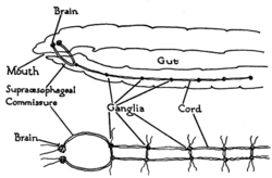
The vast majority of existing animals are bilaterians, meaning animals with left and right sides that are approximate mirror images of each other. All bilateria are thought to have descended from a common wormlike ancestor that appeared in the Cambrian period, 550–600 million years ago (Balavoine 2003). The fundamental bilaterian body form is a tube with a hollow gut cavity running from mouth to anus, and a nerve cord with an enlargement (a "ganglion") for each body segment, with an especially large ganglion at the front, called the "brain."
Even mammals, including humans, show the segmented bilaterian body plan at the level of the nervous system. The spinal cord contains a series of segmental ganglia, each giving rise to motor and sensory nerves that innervate a portion of the body surface and underlying musculature. On the limbs, the layout of the innervation pattern is complex, but on the trunk it gives rise to a series of narrow bands. The top three segments belong to the brain, giving rise to the forebrain, midbrain, and hindbrain (Ghysen 2003).
Bilaterians can be divided, based on events that occur very early in embryonic development, into two groups (superphyla) called protostomes and deuterostomes (Erwin and Davidson 2002). Deuterostomes include vertebrates as well as echinoderms, hemichordates (mainly acorn worms), and Xenoturbellidans (Bourlat et al. 2006). Protostomes, the more diverse group, include arthropods, mollusks, and numerous types of worms. There is a basic difference between the two groups in the placement of the nervous system within the body: protostomes possess a nerve cord on the ventral (usually bottom) side of the body, whereas in deuterostomes the nerve cord is on the dorsal (usually top) side. In fact, numerous aspects of the body are inverted between the two groups, including the expression patterns of several genes that show dorsal-to-ventral gradients. Most anatomists now consider that the bodies of protostomes and deuterostomes are "flipped over" with respect to each other, a hypothesis that was first proposed by Geoffroy Saint-Hilaire for insects in comparison to vertebrates. Thus insects, for example, have nerve cords that run along the ventral midline of the body, while all vertebrates have spinal cords that run along the dorsal midline (Lichtneckert and Reichert 2005).
The ventral nerve cord is a bundle of nerve fibers (typically a solid double stand or pair of cords) that runs along the longitudinal axis of some phyla of elongate invertebrates, and forms part of the invertebrate's central nervous system. In most cases, this nerve cords runs ventrally, below the gut, and connects to the cerebral ganglia. Among the phyla exhibiting ventral nerve cords are nematodes (roundworms), annelids (such as earthworms, and arthropods (such as insects and crayfish).
The spinal cord is the long, tubular structure in vertebrates that consists of a bundle of nervous tissue and support cells, connects with the brain, and extends lengthwise down the spinal cavity within the vertebral column (spine). Both the brain and the spinal cord develop from the embryonic feature known as the dorsal nerve cord.
Vertebrate Nervous System
| Peripheral | Somatic | |
| Autonomic | Sympathetic | |
| Parasympathetic | ||
| Enteric | ||
| Central | ||
The vertebrate nervous system is divided into the central nervous system and the peripheral nervous system.
The central nervous system (CNS) is composed of the brain and spinal cord and is contained within the dorsal cavity, with the brain in the cranial subcavity (the skull), and the spinal cord in the spinal cavity (within the vertebral column). The CNS is enclosed and protected by meninges, a three-layered system of membranes, including a tough, leathery outer layer called the dura mater. The brain is also protected by the skull, and the spinal cord by the vertebrae.
The peripheral nervous system (PNS) is a collective term for the nervous system structures that do not lie within the CNS. The large majority of the axon bundles called nerves are considered to belong to the PNS, even when the cell bodies of the neurons to which they belong reside within the brain or spinal cord.
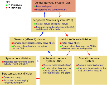
The peripheral nervous system, in turn, is commonly divided into two subsystems, the somatic nervous system and the autonomic nervous system.
The somatic nervous system (or sensory-somatic nervous system) involves nerves just under the skin, innervating skeletal muscle tissue in the skins, joints, and muscles, and serves as the sensory connection between the outside environment and the CNS. These nerves are under conscious control, but most have an automatic component, as is seen in the fact that they function even in the case of a coma (Anissimov 2007). The cell bodies of somatic sensory neurons lie in dorsal root ganglia of the spinal cord. In humans, the somatic nervous system consists of 12 pairs of cranial nerves and 31 pairs of spinal nerves (Chamberlin and Narins 2005).
The autonomic nervous system is typically presented as that portion of the peripheral nervous system that is independent of conscious control, acting involuntarily and subconsciously (reflexively), and innervating heart muscle, endocrine glands, exocrine glands, and smooth muscle (Chamberlin and Narins 2005). In sending fibers to three tissues—cardiac muscle, smooth muscle, or glandular tissue—the autonomic nervous system provides stimulation, sympathetic or parasympathetic, to control smooth muscle contraction, regulate cardiac muscle, or stimulate or inhibit glandular secretion.
The somatic nervous system always excites muscle tissue. In contrast, the autonomic nervous system can either excite or inhibit innervated tissue (Chamberlin and Narins 2005).
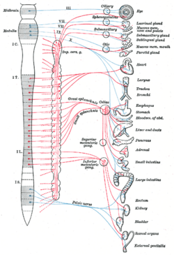
The autonomic nervous system is subdivided into the sympathetic nervous system, the parasympathetic nervous system, and the enteric nervous system. In general, the sympathetic nervous system increases activity and metabolic rate (the "fight or flight response"), while the parasympathetic nervous system slows activity and metabolic rate, returning the body to normal levels of function (the "rest and digest state") after heightened activity from sympathetic stimulation (Chamberlin and Narins 2005). The enteric nervous system innervates areas around the intestines, pancreas, and gall bladder. The role of the enteric nervous system is to manage every aspect of digestion, from the esophagus to the stomach, small intestine, and colon.
Most associated tissues and organs have nerves of both the sympathetic and the parasympathetic nervous systems. The two system can stimulate the target organs and tissues in opposite ways, such as sympathetic stimulation to increase heart rate and parasympathetic to decrease heart rate, or the sympathetic stimulation resulting in pupil dilation, and the parasympathetic in pupil constriction or narrowing (Chamberlin and Narins 2005). Or, they can both stimulate activity in concert, but in different ways, such as both increasing saliva production by salivary glands, but with sympathetic stimulation yielding viscous or thick saliva and parasympathetic yielding watery saliva. Likewise, in human reproduction, they work in concert with the parasympathetic promoting erection of genitals and the sympathetic promoting ejaculation and vaginal contractions (Campbell et al. 2008).
The vertebrate nervous system can also be divided into areas called gray matter ("grey matter" in British spelling) and white matter. Gray matter (which is only gray in preserved tissue, and is better described as pink or light brown in living tissue) contains a high proportion of cell bodies of neurons. White matter is composed mainly of myelinated axons, and takes its color from the myelin.
Invertebrate Nervous Systems
Porifera: Neural precusors
Sponges have no cells connected to each other by synaptic junctions, that is, no neurons, and therefore no nervous system. They do, however, have homologs of many genes that play key roles in synaptic function. Recent studies have shown that sponge cells express a group of proteins that cluster together to form a structure resembling a postsynaptic density (the signal-receiving part of a synapse) (Sakarya et al. 2007). However, the function of this structure is currently unclear. Although sponge cells do not show synaptic transmission, they do communicate with each other via calcium waves and other impulses, which mediate some simple actions such as whole-body contraction (Jacobs et al. 2007).
Radiata
Jellyfish, comb jellies, and related animals have diffuse nerve nets rather than a central nervous system. In most jellyfish the nerve net is spread more or less evenly across the body; in comb jellies, it is concentrated near the mouth. The nerve nets consist of sensory neurons, which pick up chemical, tactile, and visual signals; motor neurons, which can activate contractions of the body wall; and intermediate neurons, which detect patterns of activity in the sensory neurons and, in response, send signals to groups of motor neurons. In some cases, groups of intermediate neurons are clustered into discrete ganglia (Ruppert et al. 2004).
The development of the nervous system in radiata is relatively unstructured. Unlike bilaterians, radiata only have two primordial cell layers, endoderm and ectoderm. Neurons are generated from a special set of ectodermal precursor cells, which also serve as precursors for every other ectodermal cell type (Sanes et al. 2006).
Platyhelminthes, Nematoda, and Annelida
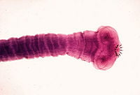
Flatworms (phylum Platyhelminthes) have a bilateral nervous system; they are the simplest animals to have one. Two cord-like nerves branch repeatedly in an array resembling a ladder. Flatworms have their sense receptors and nerves concentrated on the anterior end (cephalization). The head end of some species even has a collection of ganglia acting as a rudimentary brain to integrate signals from sensory organs, such as eyespots.
For example, planaria, a type of flatworm, have dual nerve cords running along the length of the body and merging at the tail. These nerve cords are connected by transverse nerves like the rungs of a ladder. These transverse nerves help coordinate the two sides of the animal. Two large ganglia at the head end function similar to a simple brain. Photoreceptors on the animal's eyespots provide sensory information on light and darkness.
Nematodes (roundworms, phylum Nematoda) have a simple nervous system, with a main nerve cord running along the ventral side (the "belly" side). Sensory structures at the anterior or head end are called amphids, while sensory structures at the posterior end are called phasmids.
The nervous system of the roundworm Caenorhabditis elegans has been mapped out to the cellular level. Every neuron and its cellular lineage has been recorded and most, if not all, of the neural connections are known. In this species, the nervous system is sexually dimorphic; the nervous systems of the two sexes, males and hermaphrodites, have different numbers of neurons and groups of neurons that perform sex-specific functions. In C. elegans, males have 383 neurons, while hermaphrodites have 302 neurons (Hobert 2010).
In annelids (segmented worms, phylum Annelida), the nervous system has a solid, ventral nerve cord from which lateral nerves arise in each segment. Every segment has an autonomy; however, they unite to perform as a single body for functions such as locomotion.
Arthropods
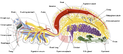
Arthropods, such as insects and crustaceans, have a nervous system made up of a series of ganglia, connected by a ventral nerve cord made up of two parallel connectives running along the length of the belly (Chapman 1998). Typically, each body segment has one ganglion on each side, though some ganglia are fused to form the brain and other large ganglia. The head segment contains the brain, also known as the supraesophageal ganglion. In the insect nervous system, the brain is anatomically divided into the protocerebrum, deutocerebrum, and tritocerebrum. Immediately behind the brain is the subesophageal ganglion, which is composed of three pairs of fused ganglia. It controls the mouthparts, the salivary glands and certain muscles. Many arthropods have well-developed sensory organs, including compound eyes for vision and antennae for olfaction and pheromone sensation. The sensory information from these organs is processed by the brain.
In insects, many neurons have cell bodies that are positioned at the edge of the brain and are electrically passive—the cell bodies serve only to provide metabolic support and do not participate in signalling. A protoplasmic fiber runs from the cell body and branches profusely, with some parts transmitting signals and other parts receiving signals. Thus, most parts of the insect brain have passive cell bodies arranged around the periphery, while the neural signal processing takes place in a tangle of protoplasmic fibers called neuropil, in the interior (Chapman 1998).
(See article on ventral nerve cord for greater detail on the arthropod nerve cord architecture.)
Mollusks
Most mollusks, such as snails and bivalves, have several groups of intercommunicating neurons called ganglia. The nervous system of the sea hare Aplysia has been extensively used in neuroscience experiments because of its simplicity and ability to learn simple associations.
The cephalopods, such as squid and octopuses, have relatively complex brains. These animals also have complex eyes. As in all invertebrates, the axons in cephalopods lack myelin, the insulator that allows fast saltatory conduction of action potentials in vertebrates. (In saltatory conduction, the action potentials do not pass continuously along the nerve, but rather "hop" from node to node in the myelin sheath along the nerve.) To achieve a high enough conduction velocity to control muscles in distal tentacles, axons in the cephalopods must have a very wide diameter in the larger species of cephalopods. For this reason, the squid giant axons were used by neuroscientists to work out the basic properties of the action potential.
ReferencesISBN links support NWE through referral fees
- Allen, N. J., and B. A. Barres. 2009. Neuroscience: Glia - more than just brain glue. Nature 457(7230): 675–7. PMID 19194443.
- Anissimov, M. 2007. How does the nervous system work? Conjecture Corporation: Wise Geek. Retrieved October 15, 2013.
- Azevedo, F. A., L. R. Carvalho, L. T. Grinberg, et al. 2009. Equal numbers of neuronal and nonneuronal cells make the human brain an isometrically scaled-up primate brain. J. Comp. Neurol. 513(5): 532–41. PMID 19226510.
- Balavoine, G. 2003. The segmented Urbilateria: A testable scenario. Int Comp Biology 43(1): 137–47. Retrieved October 15, 2013.
- Bourlat, S. J., T. Juliusdottir, C. J. Lowe, et al. 2006. Deuterostome phylogeny reveals monophyletic chordates and the new phylum Xenoturbellida. Nature 444(7115): 85–8. PMID 17051155.
- Burns, C. P. E. 2006. Altruism in nature as manifestation of divine energeia. Zygon 41(1):125-137.
- Campbell, N. A., J. B. Reece, L. A. Urry, et al. 2008. Biology, 8th edition. San Francisco: Pearson/Benjamin Cummings. ISBN 9780805368444.
- Chamberlin, S. L., and B. Narins. 2005. The Gale Encyclopedia of Neurological Disorders. Detroit: Thomson Gale. ISBN 078769150X.
- Chapman, R. F. 1998. The Insects: Structure and Function. Cambridge University Press. ISBN 9780521578905.
- Erwin, D. H., and E. H. Davidson. 2002. The last common bilaterian ancestor. Development 129(13): 3021–32. PMID 12070079.
- Finger, S. 2001. Origins of Neuroscience: A History of Explorations Into Brain Function. Oxford Univ. Press. ISBN 9780195146943.
- Ghysen, A. 2003. The origin and evolution of the nervous system. Int. J. Dev. Biol. 47(7–8): 555–62. PMID 14756331. Retrieved October 15, 2013.
- Gonzalez, R. 2012. The 4 biggest myths about the human brain. 109.com. Retrieved November 12, 2013.
- Gray, P. O. 2006. Psychology. Macmillan. ISBN 9780716776901.
- Hormuzdi, S. G, M. A. Filippov, G. Mitropoulou, et al. 2004. Electrical synapses: A dynamic signaling system that shapes the activity of neuronal networks. Biochim. Biophys. Acta 1662(1–2): 113–37. PMID 15033583.
- Hobert, O. 2010. Neurogenesis in the nematode Caenorhabditis elegans. Wormbook. Retrieved October 15, 2013.
- Jacobs, D. K., N. Nakanishi, D. Yuan, et al. 2007. Evolution of sensory structures in basal metazoa. Integr Comp Biol 47(5): 712–723. PMID 21669752. Retrieved October 15, 2013.
- Kandel, E. R., J. H. Schwartz,and T. M. Jessel (Eds.). 2000. Principles of Neural Science. McGraw-Hill Professional. ISBN 9780838577011.
- Kimball, J. W. 2011. Organization of the nervous system. Kimball's Biology Pages.
- Kimball, J. W. 2013. The human central nervous system. Kimball's Biology Pages.
- Lichtneckert, R., and H. Reichert. 2005. Insights into the urbilaterian brain: Conserved genetic patterning mechanisms in insect and vertebrate brain development. Heredity 94(5): 465–77. PMID 15770230.
- Marieb, E. N. and K. Hoehn. 2010. Human Anatomy & Physiology, 8th edition. Benjamin Cummings. ISBN 9780805395693.
- Ruppert, E. E., R. S. Fox, and R. D. Barnes. 2004. Invertebrate Zoology, 7 ed. Brooks/Cole. ISBN 0030259827.
- Sakarya, O., K. A. Armstrong, M. Adamska, et al. 2007. A post-synaptic scaffold at the origin of the animal kingdom. PLoS ONE 2(6): e506. PMID 17551586.
- Sanes, D. H., T. A. Reh, and W. A. Harris. 2006. Development of the nervous system. Academic Press. ISBN 9780126186215.
- Towle, A. 1989. Modern Biology. Austin, TX: Holt, Rinehart and Winston. ISBN 0030139198.
| Human organ systems |
|---|
| Cardiovascular system | Digestive system | Endocrine system | Immune system | Integumentary system | Lymphatic system | Muscular system | Nervous system | Skeletal system | Reproductive system | Respiratory system | Urinary system |
Credits
New World Encyclopedia writers and editors rewrote and completed the Wikipedia article in accordance with New World Encyclopedia standards. This article abides by terms of the Creative Commons CC-by-sa 3.0 License (CC-by-sa), which may be used and disseminated with proper attribution. Credit is due under the terms of this license that can reference both the New World Encyclopedia contributors and the selfless volunteer contributors of the Wikimedia Foundation. To cite this article click here for a list of acceptable citing formats.The history of earlier contributions by wikipedians is accessible to researchers here:
The history of this article since it was imported to New World Encyclopedia:
Note: Some restrictions may apply to use of individual images which are separately licensed.
