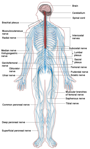
Somatic nervous system
The somatic nervous system, or voluntary nervous system, is that part of the peripheral nervous system that regulates body movement through control of skeletal (voluntary) muscles and also relates the organism with the environment through the reception of external stimuli, such as through the senses of vision, hearing, taste, and smell. The somatic nervous system controls such voluntary actions as walking and smiling through the use of efferent motor nerves, in contrast with the function of the autonomic nervous system, which largely acts independent of conscious control in innervating cardiac muscle and exocrine and endocrine glands.
Every living thing interacts with other organisms and its environment. This continuous interaction between an organism and its environment is needed for the organism to survive and grow. It is the somatic nervous system that allows individuals to receive sensory information and consciously react to environmental changes.
Overview
The somatic nervous system is one of two subsystems of the peripheral nervous system, the other being the autonomic nervous system. The autonomic nervous system is responsible for maintenance functions (metabolism, cardiovascular activity, temperature regulation, digestion) that have a reputation for being outside of conscious control. It consists of nerves in cardiac muscle, smooth muscle, and exocrine and endocrine glands. The somatic nervous system consists of cranial and spinal nerves that innervate skeletal muscle tissue and are more under voluntary control (Anissimov 2006; Towle 1989), as well as the sensory receptors.
The somatic nervous system includes all the neurons connected with muscles, skin, and sense organs. The somatic nervous system processes sensory information and controls all voluntary muscular systems within the body, with the exception of reflex arcs. The somatic nervous system consists of efferent nerves responsible for sending brain signals for muscle contraction.
Overview of human somatic nervous system
In humans, there are 31 pairs of spinal nerves and 12 pairs of cranial nerves.
The 31 pairs of spinal nerves emanate from different areas of the spinal cord and each spinal nerve has a ventral root and a dorsal root. The ventral root has motor (efferent) fibers that transmit messages from the central nervous system to the effectors, with the cell bodies of the efferent fibers found in the spinal cord gray matter. The dorsal root has sensory (afferent) fibers that carry information from the sensory receptors to the spinal cord (Adam 2001).
The 12 pairs of cranial nerves transmit information on the senses of sight, smell, balance, taste, and hearing from special sensory receptors. They also transmit information from general sensory receptors in the body, largely from the head. This information is received and processed by the central nervous system and then the response travels via the cranial nerves to the skeletal muscles to control movements in the face and throat, such as swallowing and smiling (Adam 2001).
Nerve signal transmission
The basic route of nerve signals within the efferent somatic nervous system involves a sequence that begins in the upper cell bodies of motor neurons (upper motor neurons) within the precentral gyrus (which approximates the primary motor cortex). Stimuli from the precentral gyrus are transmitted from upper motor neurons and down the corticospinal tract, via axons to control skeletal (voluntary) muscles. These stimuli are conveyed from upper motor neurons through the ventral horn of the spinal cord, and across synapses to be received by the sensory receptors of alpha motor neuron (large lower motor neurons) of the brainstem and spinal cord.
Upper motor neurons release a neurotransmitter, acetylcholine, from their axon terminal knobs, which are received by nicotinic receptors of the alpha motor neurons. In turn, alpha motor neurons relay the stimuli received down their axons via the ventral root of the spinal cord. These signals then proceed to the neuromuscular junctions of skeletal muscles.
From there, acetylcholine is released from the axon terminal knobs of alpha motor neurons and received by postsynaptic receptors (Nicotinic acetylcholine receptors) of muscles, thereby relaying the stimulus to contract muscle fibers.
In invertebrates, depending on the neurotransmitter released and the type of receptor it binds, the response in the muscle fiber could either be excitatory or inhibitory. For vertebrates, however, the response of a muscle fiber to a neurotransmitter (always acetylcholine (ACh)) can only be excitatory or, in other words, contractile.
Reflex arcs
A reflex arc is an automatic reaction that allows an organism to protect itself reflexively when an imminent danger is perceived. In response to certain stimuli, such as touching a hot surface, these reflexes are "hard wired" through the spinal cord. A reflexive impulse travels up afferent nerves, through a spinal interneuron, and back down appropriate efferent nerves.
See also
ReferencesISBN links support NWE through referral fees
- Adam. 2001. Nervous system. PNS: Somatic (voluntary) nervous system, autonomic (involuntary) nervous system. University of Tennessee Medical Center. Retrieved November 7, 2008.
- Anissimov, M. 2007. How does the nervous system work? Conjecture Corporation: Wise Geek. Retrieved November 7, 2008.
- Chamberlin, S. L., and B. Narins. 2005. The Gale Encyclopedia of Neurological Disorders. Detroit: Thomson Gale. ISBN 078769150X.
- Towle, A. 1989. Modern Biology. Austin, TX: Holt, Rinehart and Winston. ISBN 0030139198.
Credits
New World Encyclopedia writers and editors rewrote and completed the Wikipedia article in accordance with New World Encyclopedia standards. This article abides by terms of the Creative Commons CC-by-sa 3.0 License (CC-by-sa), which may be used and disseminated with proper attribution. Credit is due under the terms of this license that can reference both the New World Encyclopedia contributors and the selfless volunteer contributors of the Wikimedia Foundation. To cite this article click here for a list of acceptable citing formats.The history of earlier contributions by wikipedians is accessible to researchers here:
The history of this article since it was imported to New World Encyclopedia:
Note: Some restrictions may apply to use of individual images which are separately licensed.
