Muscle (from Latin musculus, "little mouse"), the contractile tissue of animal bodies, comprises fibers specialized to contract and effect bodily movement. In the course of embryonic development, muscle is derived from the mesodermal layer of embryonic germ cells.
Muscles exhibit complex coordination on many levels, including the filaments actin and myosin harmoniously interacting while utilizing ATP for energy to produce contraction; the successively greater scales of bundling cells to yield the mass of whole muscles; and the coordinated and appropriately scaled contraction and stretching of complementary muscle systems under the direction of the nervous system.
In vertebrates, muscle is classified as skeletal (or striated), cardiac, or smooth muscle. Its function is to produce force and cause motion, either locomotion or movement within internal organs. Much muscle contraction occurs without conscious thought and is necessary for survival, like the contraction of the heart, or peristalsis (which pushes food through the digestive system). Voluntary muscle contraction is used to move the body, and can be finely controlled, like movements of the eye, or gross movements like the quadriceps muscle of the thigh.
There are two broad types of voluntary muscle fibers, slow twitch and fast twitch. Slow twitch fibers contract for long periods of time but with little force, while fast twitch fibers contract quickly and powerfully but fatigue very rapidly.
Basic anatomy
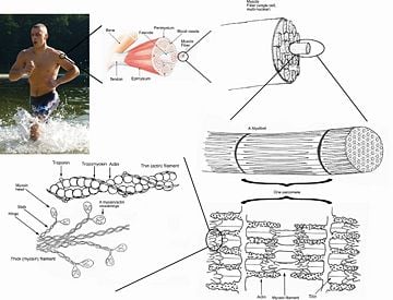
Muscle is mainly composed of muscle cells. A muscle fiber, also technically known as a myocyte, is a single cell of a muscle. Muscle fibers contain many myofibrils, the contractile units of muscles. Myofibrils are alternating bundles of thin filaments, comprising primarily actin, and thick filaments, comprising primarily the protein myosin. Myfibrils run from one end of the cell to the other. The protein complex comprising actin and myosin is sometimes referred to as "actomyosin." A skeletal muscle may contain hundreds to several thousands of myofibrils. Muscle fibers can be very short, such as 1 millimeter, to very long, such as 30 centimeters (11.8 inches).
The sarcolemma is the cell membrane enclosing each muscle fiber (muscle cell). Individual muscle fibers (including the sarcolemma) are then surrounded by endomysium, a connective tissue. Endomysium is the fine sheath of tissue that surrounds each single muscle fiber. Muscle fibers, perhaps 10 to 100 or more, are bound together by perimysium, a connective tissue, into bundles called fascicles. The bundles are then grouped together to form muscle, which is enclosed in a sheath of epimysium. That is, epimysium wraps the whole muscle. Over the layer of epimysium is fascia, a sheet of connective tissue that helps maintain form.
Muscle spindles are distributed throughout the muscles and provide sensory feedback information to the central nervous system.
Classes of muscles
There are three types of muscle:
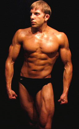
- Skeletal muscle. Skeletal muscle, also known as "striated muscle" or "voluntary muscle," is anchored (with some exceptions) by tendons to bone and is used to affect skeletal movement such as locomotion and in maintaining posture (the tongue is an example of a skeletal muscle that lacks bony supports). Skeletal muscle is also called striated because the fibers appear striped under a microscope, with alternating light and dark bands. Skeletal muscles are responsible for voluntary movement. Though postural control is generally maintained as a subconscious reflex, the muscles responsible react to conscious control like non-postural muscles. An average adult male is made up of 40-50 percent of skeletal muscle and an average adult female is made up of 30-40 percent.
- Smooth muscle. Smooth muscle, also known as "visceral muscle" or "involuntary muscle" is found within the walls of organs and structures such as the esophagus, stomach, intestines, bronchi, uterus, urethra, bladder, and blood vessels. Unlike skeletal muscle, smooth muscle is not under conscious control. It is regulated by the autonomic nervous system.
- Cardiac muscle. Cardiac muscle is also an "involuntary muscle" but is a specialized kind of muscle found only within the heart.
Cardiac and skeletal muscle are similar in that both appear to be "striated" in that they contain sarcomeres. A sarcomere is the basic functional unit of a muscle's cross-striated myofibril, the alternating bundles of filaments composed primarily of actin or myosin. In striated muscle, such as skeletal and cardiac muscle, the actin and myosin filaments each have a specific and constant length on the order of a few micrometers, far less than the length of the elongated muscle cell (a few millimeters in the case of human skeletal muscle cells). The filaments are organized into repeated subunits along the length. These subunits are called sarcomeres. A muscle cell, from a biceps, may contain 100,000 sarcomeres. The sarcomeres are what give skeletal and cardiac muscles their striated appearance. The myofibrils of smooth muscle cells are not arranged into sarcomeres.
While skeletal muscles are arranged in regular, parallel bundles, cardiac muscle connects at branching, irregular angles. Cardiac muscle is anatomically different in that the muscle fibers are typically branched like a tree branch, and connect to other cardiac muscle fibers through intercalcated discs, and form the appearance of a syncytium. Striated muscle (cardiac and skeletal) contracts and relaxes in short, intense bursts, whereas smooth muscle sustains longer or even near-permanent contractions.
On the other hand, cardiac muscle shares many properties with smooth muscle, including being controlled by the autonomic nervous system and spontaneous (automatic) contractions.
Types of skeletal muscle
Skeletal muscle is further divided into several subtypes:
- Type I, slow oxidative, slow twitch, or "red" muscle is dense with capillaries and is rich in mitochondria and myoglobin, giving the muscle tissue its characteristic red color. It can carry more oxygen and sustain aerobic activity.
- Type II, fast twitch, muscle has three major kinds that are, in order of increasing contractile speed (Larsson et al. 1991):
- Type IIa, which, like slow muscle, is aerobic, rich in mitochondria and capillaries and appears red.
- Type IIx (also known as type IId), which is less dense in mitochondria and myoglobin. This is the fastest muscle type in humans. It can contract more quickly and with a greater amount of force than oxidative muscle, but can sustain only short, anaerobic bursts of activity before muscle contraction becomes painful (often incorrectly attributed to a build-up of lactic acid). In some books and articles this muscle in humans was, confusingly, called type IIB (Smerdu et al. 1994).
- Type IIb, which is anaerobic, glycolytic, "white" muscle that is even less dense in mitochondria and myoglobin. In small animals like rodents, this is the major fast muscle type, explaining the pale color of their meat.
Skeletal muscle is arranged in discrete muscles, an example of which is the biceps brachii. It is connected by tendons to processes of the skeleton. In contrast, smooth muscle occurs at various scales in almost every organ, from the skin (in which it controls erection of body hair) to the blood vessels and digestive tract (in which it controls the caliber (opening size) of the lumen and peristalsis).
There are approximately 639 skeletal muscles in the human body. Contrary to popular belief, the number of muscle fibers cannot be increased through exercise; instead the muscle cells simply get bigger. Muscle fibers have a limited capacity for growth through hypertrophy (increase in the size of cells, while the number stays the same) and some believe they split through hyperplasia (cell division increasing the number of cells while their size stays the same) if subject to increased demand.
Physiology and contraction
The three types of muscle have significant differences. However, all three use the movement of actin against myosin to create contraction.
Muscular contraction uses adenosine triphosphate (ATP) for energy. The ATP allows, through hydrolysis, the myosin head to extend up and bind with the actin filament. The myosin head then releases after moving the actin filament in a relaxing or contracting movement by usage of ADP.
In contractile bundles, the actin-bundling protein actinin separates each filament by 40 nm. This increase in distance allows the motor protein myosin to interact with the filament, enabling deformation or contraction. In the first case, one end of myosin is bound to the cell membrane (sarcolemma) while the other end walks toward the plus end of the actin filament. This pulls the membrane into a different shape relative to the cell cortex (outer layer of cell). For contraction, the myosin molecule is usually bound to two separate filaments and both ends simultaneously walk toward their filament's plus end, sliding the actin filaments over each other. This results in the shortening, or contraction, of the actin bundle (but not the filament). This mechanism is responsible for both muscle contraction and cytokinesis, the division of one cell into two.
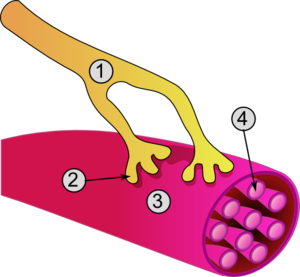
In skeletal muscle, contraction is stimulated by electrical impulses transmitted by the nerves, the motor nerves and motoneurons in particular. Cardiac and smooth muscle contractions are stimulated by internal pacemaker cells that regularly contract, and propogate contractions to other muscle cells with which they are in contact. All skeletal muscle and many smooth muscle contractions are facilitated by the neurotransmitter acetylcholine.
Muscular activity accounts for much of the body's energy consumption. All muscle cells produce ATP molecules, which are used to power the movement of the myosin heads. Muscles contain ATP in the form of creatine phosphate, which is generated from ATP and can regenerate ATP when needed with creatine kinase. Muscles also keep a storage form of glucose in the form of glycogen. Glycogen can be rapidly converted to glucose when energy is required for sustained, powerful contractions. Within the voluntary skeletal muscles, the glucose molecule is metabolized in a process called glycolysis, which produces two ATP and two lactic acid molecules in the process.
Muscle cells also contain globules of fat, which are used for energy during aerobic exercise. The aerobic energy systems take longer to produce the ATP and reach peak efficiency, and requires many more biochemical steps, but produces significantly more ATP than anaerobic glycolysis.
Cardiac muscle on the other hand, can readily consume any of the three macronutrients (protein, glucose, and fat) without a "warm up" period and always extracts the maximum ATP yield from any molecule involved. The heart and liver will also consume lactic acid produced and excreted by skeletal muscles during exercise.
Nervous control
Afferent leg
The afferent leg of the peripheral nervous system is responsible for conveying sensory information (nerve impulses) toward the central nervous system, primarily from the sense organs, like the skin.
In the muscles, the muscle spindles convey information about the degree of muscle length and stretch to the central nervous system to assist in maintaining posture and joint position. The sense of where our bodies are in space is called proprioception, the perception of body awareness. More easily demonstrated than explained, proprioception is the "unconscious" awareness of where the various regions of the body are located at any one time. This can be demonstrated by closing the eyes and waving one's hand around. Assuming proper proprioceptive function, at no time will the person lose awareness of where the hand actually is, even though it is not being detected by any of the other senses.
Several areas in the brain coordinate movement and position with the feedback information gained from proprioception. The cerebellum and red nucleus in particular continuously sample position against movement and make minor corrections to assure smooth motion.
Efferent leg
The efferent leg of the peripheral nervous system is responsible for conveying commands (nerve impulses) from the central nervous system to effectors, such as the muscles and glands. It is ultimately responsible for voluntary movement. Nerves move muscles in response to voluntary and autonomic (involuntary) signals from the brain. Deep muscles, superficial muscles, muscles of the face, and internal muscles all correspond with dedicated regions in the primary motor cortex of the brain, directly anterior to the central sulcus that divides the frontal and parietal lobes.
In addition, muscles react to reflexive nerve stimuli that do not always send signals all the way to the brain. In this case, the signal from the afferent fiber does not reach the brain, but produces the reflexive movement by direct connections with the efferent nerves in the spine. However, the majority of muscle activity is volitional, and the result of complex interactions between various areas of the brain.
Nerves that control skeletal muscles in mammals correspond with neuron groups along the primary motor cortex of the brain's cerebral cortex. Commands are routed though the basal ganglia and are modified by input from the cerebellum before being relayed through the pyramidal tract to the spinal cord and from there to the motor end plate at the muscles. Along the way, feedback loops such as that of the extrapyramidal system contribute signals to influence muscle tone and response.
Deeper muscles, such as those involved in posture, often are controlled from nuclei in the brain stem and basal ganglia.
Role in health and disease
Exercise
Exercise is often recommended as a means of improving motor skills, fitness, muscle and bone strength, and joint function. Exercise has several effects upon muscles, connective tissue, bone, and the nerves that stimulate the muscles.
Various exercises require a predominate utilization of certain muscle fibers over others. Aerobic exercise involves long, low levels of exertion in which the muscles are used at well below their maximal contraction strength for long periods of time (the most classic example being the marathon). Aerobic events, which rely primarily on the aerobic (with oxygen) system, use a higher percentage of Type I (or slow-twitch) muscle fibers, consume a mixture of fat, protein, and carbohydrates for energy, consume large amounts of oxygen, and produce little lactic acid. Anaerobic exercise involves short bursts of higher intensity contractions at a much greater percentage of their maximum contraction strength. Examples of anaerobic exercise include sprinting and weight lifting. The anaerobic energy delivery system uses predominantly Type II or fast-twitch muscle fibers, relies mainly on ATP or glucose for fuel, consumes relatively little oxygen, protein and fat, produces large amounts of lactic acid and cannot be sustained for as long a period as aerobic exercise.
The presence of lactic acid has an inhibitory effect on ATP generation within the muscle, though not producing fatigue; it can inhibit or even stop performance if the intracellular concentration becomes too high. However, long-term training causes neovascularization within the muscle, increasing the ability to move waste products out of the muscles and maintain contraction. Once moved out of muscles with high concentrations within the sarcomere, lactic acid can be used by other muscles or body tissues as a source of energy. The ability of the body to export lactic acid and use it as a source of energy depends on training level.
Delayed onset muscle soreness is the pain or discomfort often felt 24 to 76 hours after exercising and subsides generally within two to three days. Once thought to be caused by lactic acid buildup, a more recent theory is that it is caused by tiny tears in the muscle fibers caused by eccentric contraction, or unaccustomed training levels. Since lactic acid disperses fairly rapidly, it could not explain pain experienced days after exercise (Ghiasvand and Parker 2004).
Humans are genetically predisposed with a larger percentage of one type of muscle group over another. An individual born with a greater percentage of Type I muscle fibers would theoretically be more suited to endurance events, such as triathlons, distance running, and long cycling events, whereas a human born with a greater percentage of Type II muscle fibers would be more likely to excel at anaerobic events such as a 200-meter dash or weightlifting. People with high overall musculation and balanced muscle type percentage may be suited to engage in sports such as rugby or boxing.
Disease
Neuromuscular diseases are those that affect the muscles and/or their nervous control.
Symptoms or signs of muscle diseases may include weakness, spasticity (muscles are continuously contracted, causing stiffness or tightness of the muscles), myoclonus (brief, involuntary twitching of a muscle or muscle group), and myalgia (muscle pain). Diagnostic procedures that may reveal muscular disorders include testing creatine kinase levels in the blood and electromyography (measuring electrical activity in muscles). In some cases, muscle biopsy may be done to identify a myopathy (neuromuscular disease in which fibers do not function), as well as genetic testing to identify DNA abnormalities associated with specific myopathies and dystrophies.
In general, problems with nervous control can cause spasticity or paralysis, depending on the location and nature of the problem. A large proportion of neurological disorders lead to problems with movement, ranging from cerebrovascular accident (stroke) and Parkinson's disease to Creutzfeldt-Jakob disease.
A non-invasive elastography technique that measures muscle noise is undergoing experimentation to provide a way of monitoring neuromuscular disease. The sound produced by a muscle comes from the shortening of actomyosin filaments along the axis of the muscle. During contraction, the muscle shortens along its longitudinal axis and expands across the transverse axis, producing vibrations at the surface (Dume 2007).
Atrophy
There are many diseases and conditions that cause a decrease in muscle mass, known as muscle atrophy. Examples include cancer and AIDS, which induce a body wasting syndrome called cachexia. Other syndromes or conditions that can induce skeletal muscle atrophy are congestive heart disease and some diseases of the liver.
During aging, there is a gradual decrease in the ability to maintain skeletal muscle function and mass, known as sarcopenia. The exact cause of sarcopenia is unknown, but it may be due to a combination of the gradual failure in the "satellite cells," which help to regenerate skeletal muscle fibers, and a decrease in sensitivity to or the availability of critical secreted growth factors, which are necessary to maintain muscle mass and satellite cell survival. Sarcopenia is a normal aspect of aging, and is not actually a disease state.
Strength and efficiency
A display of "strength" (e.g. lifting a weight) is a result of three factors that overlap: physiological strength (muscle size, cross sectional area, available crossbridging, responses to training), neurological strength (how strong or weak is the signal that tells the muscle to contract), and mechanical strength (muscle's force angle on the lever, moment arm length, joint capabilities).
The “strongest” human muscle
Since three factors affect muscular strength simultaneously and muscles never work individually, it is unrealistic to compare strength in individual muscles and state that one is the "strongest." Accordingly, no one muscle can be named “the strongest,” but below are several muscles whose strength is noteworthy for different reasons.
- In ordinary parlance, muscular "strength" usually refers to the ability to exert a force on an external object—for example, lifting a weight. By this definition, the masseter or jaw muscle is the strongest. The 1992 Guinness Book of Records records the achievement of a bite strength of 4337 Newtons (975 lbf) for 2 seconds. What distinguishes the masseter is not anything special about the muscle itself, but its advantage in working against a much shorter lever arm than other muscles.
- If "strength" refers to the force exerted by the muscle itself, e.g., on the place where it inserts into a bone, then the strongest muscles are those with the largest cross-sectional area. This is because the tension exerted by an individual skeletal muscle fiber does not vary much. Each fiber can exert a force on the order of 0.3 micronewton. By this definition, the strongest muscle of the body is usually said to be the quadriceps femoris (front of the thigh) or the gluteus maximus (large portion of the buttocks).
- A shorter muscle will be stronger "pound for pound" (i.e., by weight) than a longer muscle. The uterus may be the strongest muscle by weight in the human body. At the time when an infant is delivered, the human uterus weighs about 1.1 kilograms (40 ounces). During childbirth, the uterus exerts 100 to 400 N (25 to 100 lbf) of downward force with each contraction.
- The external muscles of the eye are conspicuously large and strong in relation to the small size and weight of the eyeball. It is frequently said that they are "the strongest muscles for the job they have to do" and are sometimes claimed to be "100 times stronger than they need to be." However, eye movements (particularly saccades, fast movements of the eye, used on facial scanning and reading) do require high speed movements, and eye muscles are 'exercised' nightly during “rapid eye movement.”
- The unexplained statement that "the tongue is the strongest muscle in the body" appears frequently in lists of surprising facts, but it is difficult to find any definition of "strength" that would make this statement true. Note that the tongue consists of sixteen muscles, not one.
- The heart has a claim to being the muscle that performs the largest quantity of physical work in the course of a lifetime. Estimates of the power output of the human heart range from 1 to 5 watts. This is much less than the maximum power output of other muscles; for example, the quadriceps can produce over 100 watts, but only for a few minutes. The heart does its work continuously over an entire lifetime without pause, and thus does "outwork" other muscles. An output of one watt continuously for seventy years yields a total work output of two to three gigajoules.
Efficiency
The efficiency of human muscle has been measured (in the context of rowing and cycling) at 14 to 27 percent. The efficiency is defined as the ratio of mechanical work output to the total metabolic cost.
Muscle evolution
Evolutionarily, specialized forms of skeletal and cardiac muscles predated the divergence of the vertebrate/arthropod evolutionary line (Oota and Saitou 1999). This indicates that these types of muscle developed in a common ancestor sometime before 700 million years ago. Vertebrate smooth muscle (smooth muscle found in humans) was found to have evolved independently from the skeletal and cardiac muscles.
ReferencesISBN links support NWE through referral fees
- Costill, D. L., and J. H. Wilmore. 2004. Physiology of Sport and Exercise. Champaign, IL: Human Kinetics. ISBN 0736044892
- Dumé, B. 2007. “‘Muscle noise’ could reveal diseases' progression.” NewScientistTech. Retrieved June 6, 2007.
- Johnson, G. B. 2005. Biology, Visualizing Life. Holt, Rinehart, and Winston. ISBN 003016723X
- Larsson, L., L. Edstrom, B. Lindegren, L. Gorza, and S. Schiaffino. 1991. “MHC composition and enzyme-histochemical and physiological properties of a novel fast-twitch motor unit type.” The American Journal of Physiology 261(1): C93-101.
- Oota, S., and N. Saitou. 1999. “Phylogenetic relationship of muscle tissues deduced from superimposition of gene trees.” Mol. Biol. Evol. 16(6): 856-867.
- Robergs, R., F. Ghiasvand, and D. Parker. 2004. “Biochemistry of exercise-induced metabolic acidosis.” Am J Physiol Regul Integr Comp Physiol 287(3): R502-516. PMID 15308499
- Smerdu, V., I. Karsch-Mizrachi, M. Campione, L. Leinwand, and S. Schiaffino. 1994. “Type IIx myosin heavy chain transcripts are expressed in type IIb fibers of human skeletal muscle.” American Journal of Physiology 267(6): C1723-1728.
Animals : Epithelium - Connective - Muscular - Nervous
Plants : Dermal - Vascular - Ground - Meristematic
Credits
New World Encyclopedia writers and editors rewrote and completed the Wikipedia article in accordance with New World Encyclopedia standards. This article abides by terms of the Creative Commons CC-by-sa 3.0 License (CC-by-sa), which may be used and disseminated with proper attribution. Credit is due under the terms of this license that can reference both the New World Encyclopedia contributors and the selfless volunteer contributors of the Wikimedia Foundation. To cite this article click here for a list of acceptable citing formats.The history of earlier contributions by wikipedians is accessible to researchers here:
- Muscle history
- Myofibril history
- Sarcomere history
- Muscle_fiber history
- Efferent_nerve history
- Afferent_nerve history
The history of this article since it was imported to New World Encyclopedia:
Note: Some restrictions may apply to use of individual images which are separately licensed.


