NervousSystemTemporary
The nervous system is the network of specialized cells, tissues, and organs in a multicellular animal that coordinates the body's interaction with the environment, including sensing internal and external stimuli, monitoring the organs, coordinating the activity of muscles, initiating actions, and regulating behavior. All parts of the nervous system are made of nervous tissue, which conducts electrical impulses. Prominent components in a nervous system include neurons and nerves.
This major coordinating system is found in both invertebrates and vertebrates, but is most complex in vertebrate animals.
In order for an individual to grow and develop, it needs to be continuously engaged in reciprocal relationships with its environment. The nervous system is what allows that interaction with the environment. Furthermore, the ubiquity of the nervous system among multicellular organisms reflects the unity in nature.
Cephalization is a trend seen in the history of life whereby nervous tissue in more advanced organisms is concentrated toward the anterior of the body. This process culminates in a head region with sensory organs. Cephalization is intrinsically connected with a change in symmetry, accompanying the move to bilateral symmetry made in flatworms, with ocelli and auricles placed in the head region. The cephalization/bilateral symmetry combination allows animals to have sensory organs facing the direction of movement, granting a more focused assessment of the environment into which they are moving.
Neuroscience is the field of science that focuses on the study of the nervous system.
Overview
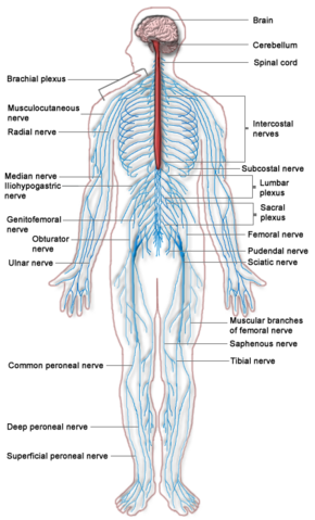
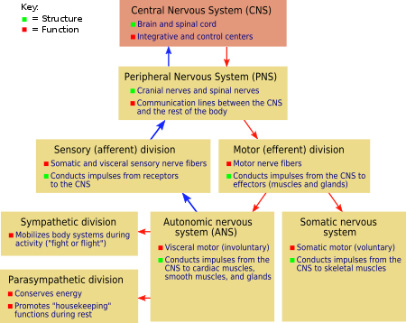
The nervous system is the part of an animal's body that coordinates the voluntary and involuntary actions of the animal and transmits signals between different parts of its body. In most types of animals it consists of two main parts, the central nervous system (CNS) and the peripheral nervous system (PNS). The CNS contains the brain and spinal cord. The PNS consists mainly of nerves, which are long fibers that connect the CNS to every other part of the body. The PNS includes motor neurons, mediating voluntary movement, the autonomic nervous system, comprising the sympathetic nervous system and the parasympathetic nervous system and regulating involuntary functions, and the enteric nervous system, a semi-independent part of the nervous system whose function is to control the gastrointestinal system.
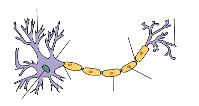
At the cellular level, the nervous system is defined by the presence of a special type of cell, called the neuron, also known as a "nerve cell". Neurons have special structures that allow them to send signals rapidly and precisely to other cells. They send these signals in the form of electrochemical waves traveling along thin fibers called axons, which cause chemicals called neurotransmitters to be released at junctions called synapses. A cell that receives a synaptic signal from a neuron may be excited, inhibited, or otherwise modulated. The connections between neurons form neural circuits that generate an organism's perception of the world and determine its behavior. Along with neurons, the nervous system contains other specialized cells called glial cells (or simply glia), which provide structural and metabolic support.
The nervous system derives its name from nerves, which are cylindrical bundles of fibers that emanate from the brain and central cord, and branch repeatedly to innervate every part of the body.[1] Nerves are large enough to have been recognized by the ancient Egyptians, Greeks, and Romans,[2] but their internal structure was not understood until it became possible to examine them using a microscope.[3] A microscopic examination shows that nerves consist primarily of the axons of neurons, along with a variety of membranes that wrap around them and segregate them into fascicles. The neurons that give rise to nerves do not lie entirely within the nerves themselves—their cell bodies reside within the brain, central cord, or peripheral ganglia.[1]
Nervous systems are found in most multicellular animals, but vary greatly in complexity.[4] The only multicellular animals that have no nervous system at all are sponges, placozoans and mesozoans, which have very simple body plans. The nervous systems of ctenophores (comb jellies) and cnidarians (e.g., anemones, hydras, corals and jellyfishes) consist of a diffuse nerve net. All other types of animals, with the exception of a few types of worms, have a nervous system containing a brain, a central cord (or two cords running in parallel), and nerves radiating from the brain and central cord. The size of the nervous system ranges from a few hundred cells in the simplest worms, to on the order of 100 billion cells in humans.
At the most basic level, the function of the nervous system is to send signals from one cell to others, or from one part of the body to others. The nervous system is susceptible to malfunction in a wide variety of ways, as a result of genetic defects, physical damage due to trauma or poison, infection, or simply aging. The medical specialty of neurology studies the causes of nervous system malfunction, and looks for interventions that can prevent it or treat it. In the peripheral nervous system, the most commonly occurring type of problem is failure of nerve conduction, which can have a variety of causes including diabetic neuropathy and demyelinating disorders such as multiple sclerosis and amyotrophic lateral sclerosis.
The need for a nervous system
Efficiencies in multicellular organisms are improved through the specialization of collections of cells to perform specific functions, such as perception, motion, ingestion, digestion, and reproduction—provided the different functions can be coordinated and the product or benefit of each functional group of cells distributed to all the other specialized groups of cells. Coordinating the activity of the specialized groups of cells is the task of the nervous system, whose level of complexity reflects the overall complexity of an organism. Examples are provided here in the worms, arthropoda, mollusca, and vertebrates.
Basic components and functions
Cells
The nervous system contains two main categories or types of cells: neurons and glial cells.
Neurons
| Neuron |
|---|
| Structure of a typical neuron |
The nervous system is defined by the presence of a special type of cell—the neuron (sometimes called "neurone" or "nerve cell").[1] Neurons can be distinguished from other cells in a number of ways, but their most fundamental property is that they communicate with other cells via synapses, which are membrane-to-membrane junctions containing molecular machinery that allows rapid transmission of signals, either electrical or chemical.[1] Many types of neuron possess an axon, a protoplasmic protrusion that can extend to distant parts of the body and make thousands of synaptic contacts.[5] Axons frequently travel through the body in bundles called nerves.
Even in the nervous system of a single species such as humans, hundreds of different types of neurons exist, with a wide variety of morphologies and functions.[5] These include sensory neurons that transmute physical stimuli such as light and sound into neural signals, and motor neurons that transmute neural signals into activation of muscles or glands; however in many species the great majority of neurons receive all of their input from other neurons and send their output to other neurons.[1]
Glial cells
Glial cells (named from the Greek for "glue") are non-neuronal cells that provide support and nutrition, maintain homeostasis, form myelin, and participate in signal transmission in the nervous system.[6] In the human brain, it is estimated that the total number of glia roughly equals the number of neurons, although the proportions vary in different brain areas.[7] Among the most important functions of glial cells are to support neurons and hold them in place; to supply nutrients to neurons; to insulate neurons electrically; to destroy pathogens and remove dead neurons; and to provide guidance cues directing the axons of neurons to their targets.[6] A very important type of glial cell (oligodendrocytes in the central nervous system, and Schwann cells in the peripheral nervous system) generates layers of a fatty substance called myelin that wraps around axons and provides electrical insulation which allows them to transmit action potentials much more rapidly and efficiently.
Overview of Bilatera, invertebrates, vertebrates
All animals more advanced than sponges have nervous systems. However, even sponges, unicellular animals, and non-animals such as slime molds have cell-to-cell signalling mechanisms that are precursors to those of neurons.[8] In radially symmetric animals such as the jellyfish and hydra, the nervous system consists of a diffuse network of isolated cells.[9] In bilaterian animals, which make up the great majority of existing species, the nervous system has a common structure that originated early in the Cambrian period, over 500 million years ago.[10]
Vertebrate Nervous Systems
| Peripheral | Somatic | |
| Autonomic | Sympathetic | |
| Parasympathetic | ||
| Enteric | ||
| Central | ||
The nervous system of vertebrate animals is often divided into the central nervous system and the peripheral nervous system. The CNS comprises the brain and spinal cord. The PNS comprises all other nerves and neurons that do not lie within the central nervous system. The large majority of what are commonly called nerves (which are actually axonal processes of nerve cells) are considered to be part of the peripheral nervous system.
The peripheral nervous system is divided further into the somatic nervous system and the autonomic nervous system.
The somatic nervous system is responsible for coordinating the body's movements, and also for receiving external stimuli. It is the system that regulates activities that are under conscious control.
The autonomic nervous system, which usually acts apart from conscious control, comprises the sympathetic division, parasympathetic division, and enteric division. The sympathetic nervous system responds to impending danger or stress, and is responsible for the increase of one's heartbeat and blood pressure, among other physiological changes, along with the sense of excitement one feels due to the increase of adrenaline in the system. The parasympathetic nervous system, on the other hand, is evident when a person is resting and feels relaxed, and is responsible for such things as the constriction of the pupil, the slowing of the heart, the dilation of the blood vessels, and the stimulation of the digestive and genitourinary systems. The role of the enteric nervous system is to manage every aspect of digestion, from the esophagus to the stomach, small intestine, and colon.
The nervous system of vertebrate animals (including humans) is divided into the central nervous system (CNS) and peripheral nervous system (PNS).[11]
The central nervous system (CNS) is the largest part, and includes the brain and spinal cord.[11] The spinal cavity contains the spinal cord, while the head contains the brain. The CNS is enclosed and protected by meninges, a three-layered system of membranes, including a tough, leathery outer layer called the dura mater. The brain is also protected by the skull, and the spinal cord by the vertebrae.
The peripheral nervous system (PNS) is a collective term for the nervous system structures that do not lie within the CNS.[12] The large majority of the axon bundles called nerves are considered to belong to the PNS, even when the cell bodies of the neurons to which they belong reside within the brain or spinal cord. The PNS is divided into somatic and visceral parts. The somatic part consists of the nerves that innervate the skin, joints, and muscles. The cell bodies of somatic sensory neurons lie in dorsal root ganglia of the spinal cord. The visceral part, also known as the autonomic nervous system, contains neurons that innervate the internal organs, blood vessels, and glands. The autonomic nervous system itself consists of two parts: the sympathetic nervous system and the parasympathetic nervous system. Some authors also include sensory neurons whose cell bodies lie in the periphery (for senses such as hearing) as part of the PNS; others, however, omit them.[13]
The vertebrate nervous system can also be divided into areas called grey matter ("gray matter" in American spelling) and white matter.[14] Grey matter (which is only grey in preserved tissue, and is better described as pink or light brown in living tissue) contains a high proportion of cell bodies of neurons. White matter is composed mainly of myelinated axons, and takes its color from the myelin. White matter includes all of the nerves, and much of the interior of the brain and spinal cord. Grey matter is found in clusters of neurons in the brain and spinal cord, and in cortical layers that line their surfaces. There is an anatomical convention that a cluster of neurons in the brain or spinal cord is called a nucleus, whereas a cluster of neurons in the periphery is called a ganglion.[15] There are, however, a few exceptions to this rule, notably including the part of the forebrain called the basal ganglia.[16]
Human nervous system
There are three essential parts of the human nervous system. These include the brain, the spine, and the nerves. The brain has three main parts that interact with the nervous system: The cerebrum, the cerebellum, and the medulla. The cerebrum's tasks include high-order thinking and learning, while the cerebellum manages learned automatic bodily functions, including walking, jumping, and running. The medulla processes simple body functions, such as breathing and digestion.
Reflex reactions occur independent of the brain with the spinal cord being the "center" of the response. Split-second reflex decisions do not involve sensory nerve impulses traveling to the brain and then back to the organ or body part. This would take too long and the nerve impulse may well arrive too late to prevent the stimulus from becoming reality. For instance, if a ball were thrown at an individual's head, the reflex to move out of the way would come from the spine, not the brain, improving reaction time. The spine is also the "highway" which passes orders from the brain to motor nerves.
There are four kinds of nerves: Motor, sensory, afferent, and interneurons. Messages carried in all nerve types travel in only one direction.
Invertebrate Nervous Systems
Porifera: Neural precusors
Worms
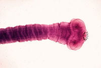
Flatworms (phylum Platyhelminthes) have a bilateral nervous system; they are the simplest animals to have one. Two cord-like nerves branch repeatedly in an array resembling a ladder. Flatworms have their sense receptors and nerves concentrated on the anterior end (cephalization). The head end of some species even has a collection of ganglia acting as a rudimentary brain to integrate signals from sensory organs, such as eyespots.
For example, planaria, a type of flatworm, have dual nerve cords running along the length of the body and merging at the tail. These nerve cords are connected by transverse nerves like the rungs of a ladder. These transverse nerves help coordinate the two sides of the animal. Two large ganglia at the head end function similar to a simple brain. Photoreceptors on the animal's eyespots provide sensory information on light and darkness.
Nematodes (roundworms, phylum Nematoda) have a simple nervous system, with a main nerve cord running along the ventral side (the "belly" side). Sensory structures at the anterior or head end are called amphids, while sensory structures at the posterior end are called phasmids.
The nervous system of the roundworm Caenorhabditis elegans has been mapped out to the cellular level. Every neuron and its cellular lineage has been recorded and most, if not all, of the neural connections are known. In this species, the nervous system is sexually dimorphic; the nervous systems of the two sexes, males and hermaphrodites, have different numbers of neurons and groups of neurons that perform sex-specific functions. In C. elegans, males have 383 neurons, while hermaphrodites have 302 neurons (Hobert 2005).
In annelids (segmented worms, phylum Annelida), the nervous system has a solid, ventral nerve cord from which lateral nerves arise in each segment. Every segment has an autonomy; however, they unite to perform as a single body for functions such as locomotion.
Arthropoda
Arthropods, such as insects and crustaceans, have a nervous system made up of a series of ganglia, connected by a ventral nerve cord which is made up of two parallel connectives running along the length of the belly. Typically, each body segment has one ganglion on each side, though some ganglia are fused to form large ganglia like the brain.
The head segment contains the brain, also known as the supraesophageal ganglion. In the insect nervous system, the brain is anatomically divided into the protocerebrum, deutocerebrum, and tritocerebrum. Immediately behind the brain is the subesophageal ganglion, which controls the mouth parts.
Many arthropods have well-developed sensory organs, including compound eyes for vision and antennae for olfactory and pheromone sensation. The sensory information from these organs is processed by the brain.
Mollusca
Most mollusks, such as snails and bivalves, have several groups of intercommunicating neurons called ganglia. The nervous system of the sea hare Aplysia has been extensively used in neuroscience experiments because of its simplicity and ability to learn simple associations.
The cephalopods, such as squid and octopuses, have relatively complex brains. These animals also have complex eyes. As in all invertebrates, the axons in cephalopods lack myelin, the insulator that allows fast saltatory conduction of action potentials in vertebrates. (In saltatory conduction, the action potentials do not pass continuously along the nerve, but rather "hop" from node to node in the myelin sheath along the nerve.) To achieve a high enough conduction velocity to control muscles in distal tentacles, axons in the cephalopods must have a very wide diameter in the larger species of cephalopods. For this reason, the squid giant axons were used by neuroscientists to work out the basic properties of the action potential.
ReferencesISBN links support NWE through referral fees
- Burns, C. P. E. 2006. Altruism in nature as manifestation of divine energeia. Zygon 41(1):125-137.
- Hobert, O. 2005. Specification of the nervous system. Wormbook. Retrieved April 28, 2007.
- Kimball, J. W. 2006.The human central nervous system. Kimball's Biology Pages. Retrieved April 28, 2007.
- Kimball, J. W. 2006. Organization of the nervous system. Kimball's Biology Pages. Retrieved April 28, 2007.
- Towle, A. 1989. Modern Biology. Austin, TX: Holt, Rinehart and Winston. ISBN 0030139198.
| Human organ systems |
|---|
| Cardiovascular system | Digestive system | Endocrine system | Immune system | Integumentary system | Lymphatic system | Muscular system | Nervous system | Skeletal system | Reproductive system | Respiratory system | Urinary system |
Credits
New World Encyclopedia writers and editors rewrote and completed the Wikipedia article in accordance with New World Encyclopedia standards. This article abides by terms of the Creative Commons CC-by-sa 3.0 License (CC-by-sa), which may be used and disseminated with proper attribution. Credit is due under the terms of this license that can reference both the New World Encyclopedia contributors and the selfless volunteer contributors of the Wikimedia Foundation. To cite this article click here for a list of acceptable citing formats.The history of earlier contributions by wikipedians is accessible to researchers here:
The history of this article since it was imported to New World Encyclopedia:
Note: Some restrictions may apply to use of individual images which are separately licensed.
- ↑ 1.0 1.1 1.2 1.3 1.4 Cite error: Invalid
<ref>tag; no text was provided for refs namedKandelCh2 - ↑ Finger S (2001). "Ch. 1: The brain in antiquity", Origins of neuroscience: a history of explorations into brain function. Oxford Univ. Press. ISBN 978-0-19-514694-3.
- ↑ Finger, pp. 43–50
- ↑ Cite error: Invalid
<ref>tag; no text was provided for refs namedColumbia - ↑ 5.0 5.1 Cite error: Invalid
<ref>tag; no text was provided for refs namedKandelCh4 - ↑ 6.0 6.1 Cite error: Invalid
<ref>tag; no text was provided for refs namedAllen2009 - ↑ Cite error: Invalid
<ref>tag; no text was provided for refs namedAzevedo - ↑ Cite error: Invalid
<ref>tag; no text was provided for refs namedSakarya - ↑ Cite error: Invalid
<ref>tag; no text was provided for refs namedRuppert - ↑ Cite error: Invalid
<ref>tag; no text was provided for refs namedBalavoine - ↑ 11.0 11.1 Cite error: Invalid
<ref>tag; no text was provided for refs namedKandelCh17 - ↑ Cite error: Invalid
<ref>tag; no text was provided for refs namedGray233 - ↑ Hubbard JI (1974). The peripheral nervous system. Plenum Press. ISBN 978-0-306-30764-5.
- ↑ Cite error: Invalid
<ref>tag; no text was provided for refs namedPurves15 - ↑ Cite error: Invalid
<ref>tag; no text was provided for refs namedDorlandsGanglion - ↑ Cite error: Invalid
<ref>tag; no text was provided for refs namedAfifi


